Friday, 3 October 2008

The wonders of auxin

I am a complete sell out. I have worked for a plant biotech company and getting funding from Gatsby (a charity with a goal, alone with others, is to get scientists interesting in plants). These people have been very nice by helping me pay rent, buy DVDs and science magazines as well as sending me to Mexico for a conference. Therefore, I thought I should give something back by educating my friends in a bit of plant biology. Don’t worry faint hearted, I will keep it nice and molecular as that is the way I like it.

After returning from my Mexico trip I realised a few things, English seems to be spoken by everyone, don’t eat food from Mexico City airport and auxin is a fascinating plant hormone and virtually everyone in the plant community agrees. Most talks there were on auxin biosynthesis, receptor function and structure, its control of shoot branching and transport around the plant. However, I do not recall all of it. Or hardy any of it as I was jet lagged with little prior knowledge of plant hormones (plus it has been over a year since the conference).

Auxin was the most interesting of all the plant signalling molecules. It has been studied for over 100 years and only today we are starting to understand how it controls plant development and cell biology. Auxin is a small molecule made from an amino acid and its most biologically active for is called IAA. Auxin controls cell elongation and division, and can promote these or stop them depending on the tissue type auxin enters. Auxin causes lateral root growth and patterns the vascular system of plants. Auxin starts shaping the plant in the embryo. What is interesting is how it regulates many of these things.

It controls gene expression by activating Auxin Response Factors (ARFs). This is done by destabilizing proteins called Aux/IAAs. Genes activated by the addition of auxin contain Auxin Response Elements (ARE) which an ARF binds to. When no auxin is present, an ARF is bound to an ARE but it is diamerized with an Aux/IAA, which represses transcription of this gene. When auxin enters the cell, it gets ride of this Aux/IAA and the ARF diamerizes with another ARF. This causes transcription of the auxin regulated gene. How does auxin cause breakdown of Aux/IAA? (I thought I would sound more intelligent if I asked a lot of questions) Auxin doesn’t have a nice receptor at the cell membrane that activates a second message or a phosphorylation pathway, no that would be to simple. In 2005 Ottoline Leyser and Stefan Kepinski (then post-doc, now lecturer at Leeds) published in Nature that TIR1 was an auxin receptor. tir1 mutants had been known to be deficient in auxin signalling for a long time but not thought to be a mutation in the receptor. TIR1 is an F-box protein, which are not famous for being receptors (until now). Normally they simply act as an E3 ubiqutin (Ub) ligase, meaning they take Ub from one protein and add it to another creating a poly-ubiquitin tag that sends a protein for degradation by the 26S proteasome. Ubiqutination and targeting to the proteasome is found animals and fungi and plays an important role in signalling, including regulation of the cell cycle! TIR1 adds Ub to the Aux/IAA when auxin enters the cell. It was assumed that auxin was perceived by some other protein in the cell and caused some modification of the Aux/IAA or TIR1 to cause this to happen, probably by phosphorylation. Now we know that auxin binds toTIR1 and acts as a molecular glue, bridging the gap between TIR1 and Aux/IAA so it can be broken down. I think this is a brilliant method. I am glad to see plants are being original and creating cool new signalling pathways instead of being just like boring old animals and fungi that love there MAPK so much they should just go along and marry it. Please see my simplified diagram of the auxin signalling pathway, it is not perfect, but who is......except maybe Colombo. But then how does auxin have very different effects on different cell types. This appears to be because different tissues expresses different ARFs and Aux/IAAs and these turn on different genes but little is known right now.


One final thing, is TIR1 the only receptor? A good question indeed. Like I said earlier, I like asking questions to make me sound smart. Other F-box proteins appear to do the same job. The same only story of redundancy. However, this style of signalling cannot explain all the effects auxin has on plants. Some auxin responses occur very quickly after auxin addition. These happen so quickly, it is unlikely proteolysis followed by transcription and translation of effectors can account for them. Things like guard cell outward K+ current up, increased cytoplasm calcium, cell wall acidification starts (to help increase cell wall expansion) and elongation growth. How can we explain auxin’s influence over these physiological changes? There is debate and investigation over what causes these things. Some are caused by changes in membrane potential and perhaps Auxin Binding Protein 1 (ABP1) has some control over some changes but I am not convinced and other features are unrelated to this. After auxin addition MAP kinase activity increase but this is unrelated to both TIR1 and ABP1. There are still a lot of unanswered questions.

Here is a short, concise review by Ottoline’s old post-doc (now at Leeds), which I found yesterday and does a far better job of introducing people to this that I have!
The anatomy of auxin perception
http://www.ncbi.nlm.nih.gov/pubmed/17876776?ordinalpos=57&itool=EntrezSystem2.PEntrez.Pubmed.Pubmed_ResultsPanel.Pubmed_DefaultReportPanel.Pubmed_RVDocSum

Another one talking a more detailed look at auxin receptors and the debate around them. I have not read it fully, sadly.
Receptors for auxin: will it all end in TIRs?
http://www.ncbi.nlm.nih.gov/sites/entrez?db=pubmed&cmd=search&term=Receptors+for+auxin%3A+will+it+all+end+in+TIRs%3F

I have not referenced here for two reasons. First, I can’t be bothered and secondly, a lot of this is from conferences or meetings I have been to. I hope you enjoyed this little rant from the plant person.

Wednesday, 1 October 2008

Rats at a rave

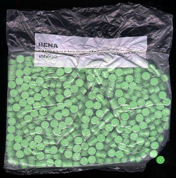
ResearchBlogging.orgThe illegal recreational drug ecstasy, also known as X, or MDMA (an abbreviation of its chemical formula), is popular in the rave scene and is used to induce euphoria and enhance the experience of dancing and loud music. The drug is considered relatively safe by the average raver due to its short term effects, despite having received publicity for various dangers identified by scientific research.

Ecstasy pills

The most widely known (undesired) effect of the drug is overexertion and sweating, leading to the ecstasy user drinking large amounts of water and being in danger of hyponatremia (the depletion of solutes from the blood plasma), which can eventually lead to cerebral edema. The drug has also been known for causing “holes in the brain”, Parkinsonian tremors, and permanent brain damage from single use, however these claims have been discredited* and retracted from the literature (1). Some research has shown that the drug may not be as toxic as once thought when used in moderation, but that does not address whether environmental effects could change the action of the drug and potentially make it more dangerous than a “traditional” toxicity study might suggest.

Research conducted in Italy and published in 2006 (2) has shown that not only does the drug enhance the rave experience for the user, but this works both ways, with the loud music physically enhancing the effect of the drug. Rats were given various doses of MDMA and treated to a surely exciting 4 hours of white noise at 95 dB (the loudest level permissible in Italian night clubs) while electrocortical activity in their brains was monitored by electroencephalography. Controls were conducted with rats treated with saline instead of MDMA, as well as both treatments without sound. ECoG monitoring was repeated over 5 days without administration of the drug or sound in order to study long term effects. The ECoG “spectrum power” was taken to be indicative of higher neural function in rats.

The data showed that a low dose of MDMA combined with sound caused decreased ECoG spectrum power significantly different from control, but no significant effect when MDMA was administered without sound. A high dose of MDMA combined with sound produced a dramatic effect that lasted for 5 days, while all other treatments had no long term effects. This powerful data shows a synergistic relationship between exposure to loud noise and the effect of MDMA on higher neural function, and suggests that higher doses only have long term effects when combined with loud noise. The authors do not suggest a possible mechanism for this effect, but they do warn that the drug may be more dangerous than commonly thought since most ecstasy users combine the drug with loud music.


* As a side note, some legislation around ecstasy was driven by the above mentioned discredited “findings”, which is disconcerting. An example is the RAVE act (3), introduced in 2002 by current Democratic VP candidate Joe Biden as part of the War On Drugs, which allows the law to shut down clubs and raves if ecstasy use is suspected to occur on their premises. The drug is still a dangerous one, but one would hope that laws would be revised after research they were based upon is discredited, which they were not.


References

1. Ronald Bailey: “The Agony of Ecstasy Research”. ReasonOnline.
2.
Michelangelo Iannone, Stefania Bulotta, Donatella Paolino, Maria Zito, Santo Gratteri, FrancescoS Costanzo, Domenicantonio Rotiroti (2006). Electrocortical effects of MDMA are potentiated by acoustic stimulation in rats BMC Neuroscience, 7 (1) DOI: 10.1186/1471-2202-7-13
3.
http://en.wikipedia.org/wiki/RAVE_Act

Environmental refugees?

As we seem to be starting a new, shorter, form of post to guarantee that someone keeps writing in this blog, I decided to follow James' example. Here I just liked to mention a short article I read at Harvard's university website. The president of Kiribati, a south Pacific island nation has given recently a lecture at Harvard where he has presented his plan preparing his country for the eventuality (or should we call it certainty) of extinction. Apparently, with the sea-levels planned to raise by 1 meter in the next century due to climate change, the islands are starting to run out of space, and to eventually leave the islands for good seems to be the only option. However, the president seems to want to avoid creating what he describes as 'environmental refugees', a new word that we will perhaps become more and more familiar with in the future, as climate change starts having 'real' consequences that we cannot pretend to ignore anymore.

Read the short article here: http://www.news.harvard.edu/gazette/2008/09.25/13-kiribati.html

Sunday, 28 September 2008

Facts about genes

As I am the saddest of the three musket-geneticists by far I have decided to post something. I bought a book called ‘a short guide to the Human Genome’. The booked is aimed at people with a background in genetics. Good for lecturers to liven up lectures with facts. The book is a series of questions on various parts of molecular biology and ‘omics’ with short, one page long, answers. These are interesting questions but difficult to find the answers to them without a lot and more importantly, knowing where to look. So here I will look at a few of the more interesting and simple questions.

How many genes are there?
22,740 predicted and known genes. All genes minus predicted transcripts gives us only 18,357.
What is the typical size of a gene?
The median sixe is 16,995 nucleotides.
Which are the largest genes?
CNTNAP2 is 2.3 Mb (remember the genome of E. coli is only 4.63 Mb) and it generates a mRNA of 9.9 Kb. DMD which makes dystrophin is the second largest gene at 2.22 Mb making an mRNA of 14.1 Kb.
Largest proteins?
TTN gene which makes titin, which is 33,423 amino acid residues long. Mucin 16 is the second largest at 14,507 amino acid residues.
How much of the genome is made up of transposable elements?
45%: SINEs 13%, LINEs 21%, LTRs 8% and DDNA transposons 3%.
How many pseudogenes are there?
There are ~5000 pseudogenes with a median size of 1200 nucleotides.
The books seems good but I would suggest you borrow it off me rather than buy a copy.

Sunday, 10 August 2008

Cross presentation: presenting unexpected antigens to the cell-mediated immune response.

Please cast your minds back to basic immunology. I am sure you remember there is the innate immune response (boring) and the acquired/adaptive immune response (very interesting). Perhaps the most interesting part of it is the gene re-arrangement to generate antibodies and T cell receptors (TCRs). But this re-arrangement is random; how can they tell self from non-self? The very basic explanation is seems to be that antigen (Ag) presenting cells (APCs) shows lymphocytes these Ag in the local draining lymph nodes when the body knows it is ill (ie the innate immune system is acting up). (Sorry for this long introduction to the topic but I need to remind myself of this while I type on the train myself.)

A problem comes when we remember a bit of basic immunology. Peptide Ag presented on MHC class II molecules to CD4+ T cells (T helper cells) are taken up by APCs and are exogenous Ag. However, peptides shown on MHC class I to CD8+ T cells are generated from endogenous proteins. So how can a virus that infects only the liver like hepatitis or the respiratory system (like rhinovirus or flu) show their peptide Ag to naive CD8+ T cells (cytotoxic T cells or CTLs for short) which live in the lymph organs. Unless all viruses also affect the APCs as well then it seems impossible! Let’s forget how ridicules that idea is first and remember CTLs also attack tumour cells expressing mutant proteins. Cross presentation appears to be the answer. Somehow, APCs such as dendritic cells (DC) take up proteins from other cells and process these in a manner that means they are treated like endogenous proteins and therefore processed to be peptide Ag loaded onto MHC class I. This is called cross presentation, when CTLs are activated this way they are cross primed. The fine details of this appear to be missing but a lot of evidence is mounting that shows this must be the case. Other than this I can see no other way apart from the naive CTLs circulating the body and activating their; but no evidence has been found for this that I can see (I am pretty sure this also flies in the face of most accepted ideas in immunology). There are four ways for DC to capture extracellular proteins; (i) endocytosis (ii) pinocytosis (cell drinking) (iii) phagocytosis and (iv) macropinocytosis. For example, phagocytosis can be the uptake of a bacterium or cellular debris such as an apoptotic body. When cells are signalled to undergo apoptosis the dying cell starts to bleb and releases intact fragments of the cell called apoptotic bodies that express ‘eat-me’ signals. It is not a huge leap of the imagination to think this could be a major way tumour proteins are presented to naive CTLs.

So how do these exogenous proteins within sub-cellular compartments such as the endosome or phagosome get loaded onto the MHC class I molecules? It is a good question and various routes have been suggested and evidence for some of these have been found. In DC exogenous proteins have been shown to be exported into the cytosol before they are degraded in the lysosome. This means they can be substrate for the proteasome and broken down to short peptides to be exported across the ER membrane by TAP. When here they can bind to the peptide binding cleft of MHC class I molecules and move to the cell surface like normal endogenous proteins in the classical presentation pathway. Another pathway acts in the endosome. This TAP/proteasome independent pathway uses cathepsin S to generate some peptide Ag. It is unclear whether other peptides here also act but currently appears unlikely. What I find amazing is how this protease produces peptides of the correct size of 8 or 9 amino acids residues long to load onto MHC class I molecules. This pathway even produces the same peptide Ag as the much more complex proteasome based pathway. When both pathways are knocked out most of the cross-presentation in vivo is lost.
Interestingly, tolerance to an Ag can be generated using these mechanisms when no stimulator of the immune system is present, and this has been called cross tolerance. It is now clear cross presentation is not some strange phenomenon like originally thought when discovered but is a key part of the immune system. When you knockout these pathways for cross-presentation none occurs. The next step is to manipulate this with vaccines to stimulate the cell-mediated immune response. Often only antibodies are produced because the antigens are not cross-presented.

A short and sweet review:
Brode and Macary (2004) Cross-presentation: dendritic cells and macrophages bite off more than they can chew!

A full and great review:
Rock and Shen (2005) Cross-presentation: underlying mechanisms and role in immune surveillance.