Friday 17 October 2008

Kinetoplastid DNA

ResearchBlogging.orgIt seems to be that time of the year in which we post about something to do with our posts, as these are the topics we are supposed to be reading about now. Therefore, a post on parasites. That’s where the connection with my project finishes, but anyway.

As you might know, tryponosomatids are organisms from the family Troponosomatidae, which include some pretty nasty unicellular parasites such as Trypanosoma brucei, causer for African sleeping disease, and Leishmania, causer of leishmaniasis. As other higher eukaryotes, these parasites are characterised by two genomes: a nuclear genome and a mitochondrion genome, 10-30% of all the cell DNA, situated in the kinetoplast (the name given to the mitochondrion in these organisms).

Fig.1 A- Schematic minicircle organization. B- in vivo network organization, seen sideways.

(Adapted from reference 1)

So far nothing extraordinary. Things get interesting, when one looks at the structure of the kinetoplast DNA (kDNA) in these species. The kDNA is constituted by thousands of small DNA circles, the minicircles, and a few dozen of large DNA circles, the maxicircles. These are interlocked, as in a chain mail of medieval armour. Each circle is interlocked with 3 other neighbouring circles. The minicircles and maxicircles are stretched into a disk-shaped structure, situated near to the flagellar basal body (Fig.1). The maxicircles are those more similar to normal mitochondrion genome. They include rRNAs and genes encoding proteins associated with the respiratory processes that takes place in this organelle. However, the maxicircles mRNA requires extensive editing, namely the introduction or deletion of uridylate. The minicircles encode the guide RNAs used as templates for this editing. Since there are many types of editing required, as well as maxicircles with different sequences, there is the need for the thousand minicircles with different sequences. Seems a bit of a wasteful system, but what do I know!

More interesting is to think of the replication process. The minicircles must stop being interlocked, replicated, interlocked again, and in the end originate two new networks of thousands of minicircles that separate into the two new daughter cells. Let us look at the different steps, following an individual minicircle (Fig.2):

Fig.2 kDNA replication model. (From reference 2)

1- A minicircle must first be released from the network into the so-called kinetoflagellar zone (KTZ), where several proteins involved in the process exist. Here the unidirectional replication of the circle occurs, although the details of this process are not well known.

2- The two daughter minicircles move to opposite antipodal sites of the circle, where there are two protein assemblies. Here a variety of reactions occur: the RNA primers are removed, the gaps between Okazaki fragments are filled by DNA pol, and nick as sealed by DNA ligase. An important characteristic, however, is that the minicircles maintain at least one gap in their structure. This is thought to be a self-check feature of this system, as a way to guarantee that no minicircle is replicated twice.

3- Minicircles are attached by topoisomerase II to the network periphery adjacent to the antipodal sites.

The way the minicircles are attached to the disk varies with the species of trypanosomatid. In T.brucei, for example, the minicircles accumulate at the network poles. In T.cruzi, and C.fasciculata, the minicircles seem to be uniformly attached around the periphery of the disk, creating a ring of new minicircles, of increased thickness. How this uniform attachment is done is not certain, but it is though that either the antipodal protein complexes or the disk itself must rotate! (Fig.3)

Fig.3 Replication in C.fasciculata. New minicircles have been labelled with fluorescent nucleotides. The arrangement of the new minicircles around the periphery of the disk is obvious (from reference 2).


4- Regardless of the method, new minicircles are attached to the disk, and the valence must increase to 4 to 6 attachments per minicircle, in order to ensure twice as many minicircles in the same space (the mitochondrion membrane has not doubled at this point). When the space increases, topoisomerase II ensures that valence returns to 3.

5- Finally, the nicks are repaired and the network splits into 2, although how exactly this division occurs is not well known. Most importantly, it is not known for sure how to guarantee that the two daughter networks have the exactly same minicircles. Considering their importance in maxicircle mRNA editing, the loss of one minicircle could have dramatic consequences.

There is much to be explained in this process, namely identify exactly how the minicircles are moved to the different antipodal sites, or the details of individual minicircle replication, but such understanding will require the identification of the proteins involved. Considering the complexity of the process, many must be involved, but only a few have been identified so far.

This is probably not life-changing research, but it is surely interesting. It is important to remind ourselves once in a while that though we learn the textbook pathways and processes, Nature seems to like using hundreds of alternatives. Just to make scientists lives harder, I assume!


Liu, B., Liu, Y., Motyka, S., Agbo, E., & Englund, P. (2005). Fellowship of the rings: the replication of kinetoplast DNA Trends in Parasitology, 21 (8), 363-369 DOI: 10.1016/j.pt.2005.06.008

(2) Smith D. and Parsons M. (1996). Molecular Biology of Parasitic Protozoa, Kinetoplast DNA: Structure and Replication, Oxford University Press, USA

Sunday 12 October 2008

Apical dominance, MAX and my project

One of the most important things the plant hormone auxin does is play an important role in is apical dominance. This is where the Shoot Apical Meristem (SAM), the growing part of the above ground half of the plant, produces a signal that inhibits growth from other auxiliary meristems. The SAM and auxiliary meristems contain the plant’s ‘stem cells’ and generate new leaves, stems and flowers. They both have the same ability to generate new organs. When the plant grows and leaves emerge from the meristem, an auxiliary meristem is left behind with the leaf, which may or may not grow out, just above where the leaf meets the stem. For years it was known auxin from the SAM inhibited their outgrowth. Auxin is produced in the SAM and moved down the plant and when it is stopped, the auxiliary meristem can grow out. The best person to tell you this is a gardener. When pruning, they remove the SAM on a plant, like a rose bush, to allow dormant growing parts of the plant grow out and make it bushier. It is also a pain for, let’s say for example, tobacco farmers. They ‘top’ tobacco plants so it does not grow up and make the leaves fatter. However, this stops auxin travelling down the stem and auxiliary meristems grow out. The addition of unpleasant chemicals is used to solve this problem.


Auxin does not travel up into the auxiliary meristem, so how does auxin inhibit bud outgrowth? A second signal must be involved. This ‘second messenger’ is not an easy thing to understand. It turns out to be a complex interplay of plant hormones that I will try to explain now control bud outgrowth. I will start by explaining the players involved. The hormone cytokinin (Ck) often plays the opposite role to auxin in plant grow. When it is applied to an auxiliary meristem it will grow out. Ck is generated both in the roots and locally in the stem but it is unclear which is more relevant to shoot branching (I think it varies from species to species). The auxin signalling pathway does regulate Ck synthesis but this is not the whole story. Auxin signalling pathway mutants do show some increase in shoot branching but it is lower than those affected by mutations in MAX genes (see below) and the effects of these are additive to auxin signalling.


Mutant screens reviled the MAX genes. max mutants have increased shoot branching. To cut a long story short, the MAX pathway does not produce the inhibitory signal but helps auxin regulate shoot-branching. MAX3 and 4 produce enzymes that alter a caratinoid. MAX1 is a P450 that acts downstream of MAX3 and 4 and alters the chemical further, producing the MAX-dependent hormone (more on the identity below). MAX2 does not produce the graft-transmissible signal but helps perceives the MAX-dependent hormone. In fact, it is an F-box protein like TIR1 from my post on auxin signalling. However, I think it is unlikely to bind the MAX-dependent hormone directly as TIR1 does auxin. I suppose this cannot be looked at until the hormone has been characterised better. MAX2 is also involved with leaf senescence and some features of light perception but its roles in these are not very well understood either.

The Leyser lab has been working on finding the identity of the MAX-dependent hormone but two papers published in the same September issue of Nature (just after my subscription ended!) suggest the identity in pea and rice. Ottoline has just written a short review on them (below). They found a chemical, called strigolactones, was involved. Previously shown to be involved with germination, formation of mycorrhiza with fungi and growth of parasitic plants, now it appears they are a key regulator of shoot branching. The exact identity of the biological active strigolactone is still yet to be found.

The MAX pathway’s mode of action is through limiting auxin transport. This work was published with a PhD student at York as the lead author in the Leyser lab (he was a very well known blue coat). Auxin is transported from the SAM to the roots. To move in and out of cells, auxin needs proteins to transport it. One important class are the PIN proteins. They can become localised to a particular part in a plant cell, such as the basal side, to ensure auxin only moves in one direction. This is very important in creating a vascular system in the plant. First of all, auxin is made by the auxiliary meristems but it is their ability to transport auxin out that allows them to grow out. It is best to imagine the auxin transport network as roads. Auxin (the cars) leaves the SAM and moves down the stem via PIN proteins (lanes on a motor way). In a normal plant, not all lanes are open. So auxin from the SAM fills most of the PIN proteins and only a little auxin from the buds can enter, letting some grow out. max mutants have an increase transport capacity because more PIN proteins are present. This is like opening extra lanes on a motor way so auxin from all the buds can enter and move freely, letting them grow out! Integrating the actions of all hormones leads to something like this: auxin moves down the plant and its movement down is limited by the MAX-dependent hormone from the roots. This limits the amount of auxin auxiliary merisetems can transport out, so large amounts of auxin accumulate in these buds. This (somehow) inhibits Ck production to limit growth of the bud. The integration of multiple hormones has been described as the brain of plants.

The reason max mutants and auxin signalling double mutants have an additive effect is because auxin works both through its classical signalling pathway but also through auxin transport. This is where, for me, things get confusing. How does auxin in the bud know not to be exported and form vascular tissue to connect to the main flow in the stem? This has been a long standing question, not for shoot branching but for the formation of a vascular network, yet there is no answer (or good one at least).

My final year project is in the Leyser lab, for what I hope are obvious reasons now. They have done lots of great work. A modifier of the max1 phenotype was found in a mutants screen and mapped to a location on chromosome 1. My job is to widdle the candidate genes down from around 20 to one! I don’t want to give too much away about the project on here but the mutation alters the max1 phenotype and has its own slight developmental phenotype. Hopefully understanding it will help in the long term goal of understanding plant development. I doubt it will have a direct role synthesis or degradation of the MAX-dependent hormone but I believe it will affect downstream events. I hope this has been enlighting.

Here is a very good review and explains these things better than I can do by someone in the Leyser lab:
http://www.ncbi.nlm.nih.gov/pubmed/17728300?ordinalpos=4&itool=EntrezSystem2.PEntrez.Pubmed.Pubmed_ResultsPanel.Pubmed_DefaultReportPanel.Pubmed_RVDocSum

Here is an ahead of print short review on the identity of the MAX-dependent hormone by Ottoline:
http://www.ncbi.nlm.nih.gov/pubmed/18804430?ordinalpos=1&itool=EntrezSystem2.PEntrez.Pubmed.Pubmed_ResultsPanel.Pubmed_DefaultReportPanel.Pubmed_RVDocSum

Friday 3 October 2008

IgNobels 2008

'For achievements that first make people LAUGH and then make them THINK'

As I seem to be spending all my holidays memorizing hard words in english (Melly will know what I mean) I actually haven't had the time to read anything 'scientific' like you guys. I had, however, time to have a look to what Nature regards as, and I agree, 'the highlight of the scientific calendar'. Yes, my friends, the Ig Nobels are out again. As you will remember, I wrote a post last year when I realized that these amazing prizes existed, and have been eagerly waiting for this year's winners. So, below, enjoy the list of the IgNobel winners 2008!

NUTRITION

Massimiliano Zampini of the University of Trento, Italy and Charles Spence of OxfordUniversity,UK, for electronically modifying the sound of a potato chip to make the person chewing the chip believe it to be crisper and fresher than it really is.

PEACE
The Swiss Federal Ethics Committee on Non-Human Biotechnology (ECNH) and the citizens of Switzerland for adopting the legal principle that plants have dignity.

ARCHEOLOGY Astolfo G. Mello Araujo and José Carlos Marcelino of Universidade de São Paulo, Brazil, for measuring how the course of history, or at least the contents of an archaeological dig site, can be scrambled by the actions of a live armadillo.

BIOLOGY
Marie-Christine Cadiergues, Christel Joubert,, and Michel Franc of Ecole Nationale Veterinaire de Toulouse, France for discovering that the fleas that live on a dog can jump higher than the fleas that live on a cat.

MEDICINE
Dan Ariely of Duke University, USA, for demonstrating that high-priced fake medicine is more effective than low-priced fake medicine. (a study on placebo effects)

COGNITIVE SCIENCE
Toshiyuki Nakagaki of Hokkaido University, Japan, Hiroyasu Yamada of Nagoya, Japan, Ryo Kobayashi of Hiroshima University, Atsushi Tero of Presto JST, Akio Ishiguro of Tohoku University, and Ágotá Tóth of the University of Szeged, Hungary, for discovering that slime molds can solve puzzles.

ECONOMICS

Geoffrey Miller, Joshua Tybur and Brent Jordan of the University of New Mexico, USA, for discovering that a professional lap dancer's ovulatory cycle affects her tip earnings.

PHYSICS
Dorian Raymer of the Ocean Observatories Initiative at Scripps Institution of Oceanography, USA, and Douglas Smith of the University of California, San Diego, USA, for proving mathematically that heaps of string or hair or almost anything else will inevitably tangle themselves up in knots.

CHEMISTRY Sharee A. Umpierre of the University of Puerto Rico, Joseph A. Hill of The Fertility Centers of New England (USA), Deborah J. Anderson of Boston University School of Medicine and Harvard Medical School (USA), for discovering that Coca-Cola is an effective spermicide, and to Chuang-Ye Hong of Taipei Medical University (Taiwan), C.C. Shieh, P. Wu, and B.N. Chiang (all of Taiwan) for discovering that it is not.

LITERATURE
David Sims of Cass Business School. London, UK, for his lovingly written study "You Bastard: A Narrative Exploration of the Experience of Indignation within Organizations."

Hehe, as usual, delicious to read... For more information on the Ig Nobels or this year's ceremony (I actually don't know yet what's this year's topic. Last year it was chicken and involved dressing proper Nobel laureates in egg outfits), see the link below

http://improbable.com/ig/winners/#ig2008

One of last year's winners, Dan Meyer demonstrates his skills, after winning the 2007 IgNobel for medicine in collaboration with Brian Witcombe, for their study on sword swallowing and its side-effects

The wonders of auxin

I am a complete sell out. I have worked for a plant biotech company and getting funding from Gatsby (a charity with a goal, alone with others, is to get scientists interesting in plants). These people have been very nice by helping me pay rent, buy DVDs and science magazines as well as sending me to Mexico for a conference. Therefore, I thought I should give something back by educating my friends in a bit of plant biology. Don’t worry faint hearted, I will keep it nice and molecular as that is the way I like it.

After returning from my Mexico trip I realised a few things, English seems to be spoken by everyone, don’t eat food from Mexico City airport and auxin is a fascinating plant hormone and virtually everyone in the plant community agrees. Most talks there were on auxin biosynthesis, receptor function and structure, its control of shoot branching and transport around the plant. However, I do not recall all of it. Or hardy any of it as I was jet lagged with little prior knowledge of plant hormones (plus it has been over a year since the conference).

Auxin was the most interesting of all the plant signalling molecules. It has been studied for over 100 years and only today we are starting to understand how it controls plant development and cell biology. Auxin is a small molecule made from an amino acid and its most biologically active for is called IAA. Auxin controls cell elongation and division, and can promote these or stop them depending on the tissue type auxin enters. Auxin causes lateral root growth and patterns the vascular system of plants. Auxin starts shaping the plant in the embryo. What is interesting is how it regulates many of these things.

It controls gene expression by activating Auxin Response Factors (ARFs). This is done by destabilizing proteins called Aux/IAAs. Genes activated by the addition of auxin contain Auxin Response Elements (ARE) which an ARF binds to. When no auxin is present, an ARF is bound to an ARE but it is diamerized with an Aux/IAA, which represses transcription of this gene. When auxin enters the cell, it gets ride of this Aux/IAA and the ARF diamerizes with another ARF. This causes transcription of the auxin regulated gene. How does auxin cause breakdown of Aux/IAA? (I thought I would sound more intelligent if I asked a lot of questions) Auxin doesn’t have a nice receptor at the cell membrane that activates a second message or a phosphorylation pathway, no that would be to simple. In 2005 Ottoline Leyser and Stefan Kepinski (then post-doc, now lecturer at Leeds) published in Nature that TIR1 was an auxin receptor. tir1 mutants had been known to be deficient in auxin signalling for a long time but not thought to be a mutation in the receptor. TIR1 is an F-box protein, which are not famous for being receptors (until now). Normally they simply act as an E3 ubiqutin (Ub) ligase, meaning they take Ub from one protein and add it to another creating a poly-ubiquitin tag that sends a protein for degradation by the 26S proteasome. Ubiqutination and targeting to the proteasome is found animals and fungi and plays an important role in signalling, including regulation of the cell cycle! TIR1 adds Ub to the Aux/IAA when auxin enters the cell. It was assumed that auxin was perceived by some other protein in the cell and caused some modification of the Aux/IAA or TIR1 to cause this to happen, probably by phosphorylation. Now we know that auxin binds toTIR1 and acts as a molecular glue, bridging the gap between TIR1 and Aux/IAA so it can be broken down. I think this is a brilliant method. I am glad to see plants are being original and creating cool new signalling pathways instead of being just like boring old animals and fungi that love there MAPK so much they should just go along and marry it. Please see my simplified diagram of the auxin signalling pathway, it is not perfect, but who is......except maybe Colombo. But then how does auxin have very different effects on different cell types. This appears to be because different tissues expresses different ARFs and Aux/IAAs and these turn on different genes but little is known right now.


One final thing, is TIR1 the only receptor? A good question indeed. Like I said earlier, I like asking questions to make me sound smart. Other F-box proteins appear to do the same job. The same only story of redundancy. However, this style of signalling cannot explain all the effects auxin has on plants. Some auxin responses occur very quickly after auxin addition. These happen so quickly, it is unlikely proteolysis followed by transcription and translation of effectors can account for them. Things like guard cell outward K+ current up, increased cytoplasm calcium, cell wall acidification starts (to help increase cell wall expansion) and elongation growth. How can we explain auxin’s influence over these physiological changes? There is debate and investigation over what causes these things. Some are caused by changes in membrane potential and perhaps Auxin Binding Protein 1 (ABP1) has some control over some changes but I am not convinced and other features are unrelated to this. After auxin addition MAP kinase activity increase but this is unrelated to both TIR1 and ABP1. There are still a lot of unanswered questions.

Here is a short, concise review by Ottoline’s old post-doc (now at Leeds), which I found yesterday and does a far better job of introducing people to this that I have!
The anatomy of auxin perception
http://www.ncbi.nlm.nih.gov/pubmed/17876776?ordinalpos=57&itool=EntrezSystem2.PEntrez.Pubmed.Pubmed_ResultsPanel.Pubmed_DefaultReportPanel.Pubmed_RVDocSum

Another one talking a more detailed look at auxin receptors and the debate around them. I have not read it fully, sadly.
Receptors for auxin: will it all end in TIRs?
http://www.ncbi.nlm.nih.gov/sites/entrez?db=pubmed&cmd=search&term=Receptors+for+auxin%3A+will+it+all+end+in+TIRs%3F

I have not referenced here for two reasons. First, I can’t be bothered and secondly, a lot of this is from conferences or meetings I have been to. I hope you enjoyed this little rant from the plant person.

Wednesday 1 October 2008

Rats at a rave

ResearchBlogging.orgThe illegal recreational drug ecstasy, also known as X, or MDMA (an abbreviation of its chemical formula), is popular in the rave scene and is used to induce euphoria and enhance the experience of dancing and loud music. The drug is considered relatively safe by the average raver due to its short term effects, despite having received publicity for various dangers identified by scientific research.

Ecstasy pills

The most widely known (undesired) effect of the drug is overexertion and sweating, leading to the ecstasy user drinking large amounts of water and being in danger of hyponatremia (the depletion of solutes from the blood plasma), which can eventually lead to cerebral edema. The drug has also been known for causing “holes in the brain”, Parkinsonian tremors, and permanent brain damage from single use, however these claims have been discredited* and retracted from the literature (1). Some research has shown that the drug may not be as toxic as once thought when used in moderation, but that does not address whether environmental effects could change the action of the drug and potentially make it more dangerous than a “traditional” toxicity study might suggest.

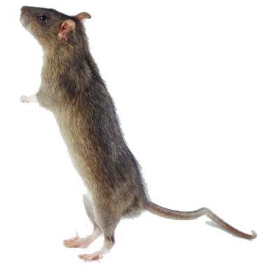
Research conducted in Italy and published in 2006 (2) has shown that not only does the drug enhance the rave experience for the user, but this works both ways, with the loud music physically enhancing the effect of the drug. Rats were given various doses of MDMA and treated to a surely exciting 4 hours of white noise at 95 dB (the loudest level permissible in Italian night clubs) while electrocortical activity in their brains was monitored by electroencephalography. Controls were conducted with rats treated with saline instead of MDMA, as well as both treatments without sound. ECoG monitoring was repeated over 5 days without administration of the drug or sound in order to study long term effects. The ECoG “spectrum power” was taken to be indicative of higher neural function in rats.

The data showed that a low dose of MDMA combined with sound caused decreased ECoG spectrum power significantly different from control, but no significant effect when MDMA was administered without sound. A high dose of MDMA combined with sound produced a dramatic effect that lasted for 5 days, while all other treatments had no long term effects. This powerful data shows a synergistic relationship between exposure to loud noise and the effect of MDMA on higher neural function, and suggests that higher doses only have long term effects when combined with loud noise. The authors do not suggest a possible mechanism for this effect, but they do warn that the drug may be more dangerous than commonly thought since most ecstasy users combine the drug with loud music.


* As a side note, some legislation around ecstasy was driven by the above mentioned discredited “findings”, which is disconcerting. An example is the RAVE act (3), introduced in 2002 by current Democratic VP candidate Joe Biden as part of the War On Drugs, which allows the law to shut down clubs and raves if ecstasy use is suspected to occur on their premises. The drug is still a dangerous one, but one would hope that laws would be revised after research they were based upon is discredited, which they were not.


References

1. Ronald Bailey: “The Agony of Ecstasy Research”. ReasonOnline.
2.
Michelangelo Iannone, Stefania Bulotta, Donatella Paolino, Maria Zito, Santo Gratteri, FrancescoS Costanzo, Domenicantonio Rotiroti (2006). Electrocortical effects of MDMA are potentiated by acoustic stimulation in rats BMC Neuroscience, 7 (1) DOI: 10.1186/1471-2202-7-13
3.
http://en.wikipedia.org/wiki/RAVE_Act

Environmental refugees?

As we seem to be starting a new, shorter, form of post to guarantee that someone keeps writing in this blog, I decided to follow James' example. Here I just liked to mention a short article I read at Harvard's university website. The president of Kiribati, a south Pacific island nation has given recently a lecture at Harvard where he has presented his plan preparing his country for the eventuality (or should we call it certainty) of extinction. Apparently, with the sea-levels planned to raise by 1 meter in the next century due to climate change, the islands are starting to run out of space, and to eventually leave the islands for good seems to be the only option. However, the president seems to want to avoid creating what he describes as 'environmental refugees', a new word that we will perhaps become more and more familiar with in the future, as climate change starts having 'real' consequences that we cannot pretend to ignore anymore.

Read the short article here: http://www.news.harvard.edu/gazette/2008/09.25/13-kiribati.html